| T O P I C R E V I E W |
| Francesco |
Posted - 15/03/2014 : 11:19:26
The shape of the genitalia is oft an important differential character for many groups.
Nonetheless, the extraction may become an insurmountable problem for experienced entomologists as well, who never tried to do it.
The description of this technique is sometimes long and difficult to be understood; thus, I decided to insert a description rich in pictures.
In this first topic, I will deal with the extraction of members of the subfamily Lepturinae, with which I suggest to start to practice.
In this group, in fact, the abdomen is fairly mobile,favoring the manipulation of the beetle.
Many thanks to my wife Otilia for having helped me take these pictures.
|
| 12 L A T E S T R E P L I E S (Newest First) |
| kbdesai |
Posted - 18/02/2016 : 18:07:09
Absolutely brilliant work Francesco... |
| nalslan |
Posted - 17/09/2015 : 17:22:21
Very nice tutorial!
I wonder what is your thought on using KOH solution to resolve muscles and membranes for the extraction? |
| amitavaeco |
Posted - 17/08/2015 : 18:31:01
Excellent work Sir |
| Xavier |
Posted - 16/05/2014 : 07:08:04
Wouah  Francesco ! Quel exposé ! Francesco ! Quel exposé !
Merci ŕ Otilia  |
| Francesco |
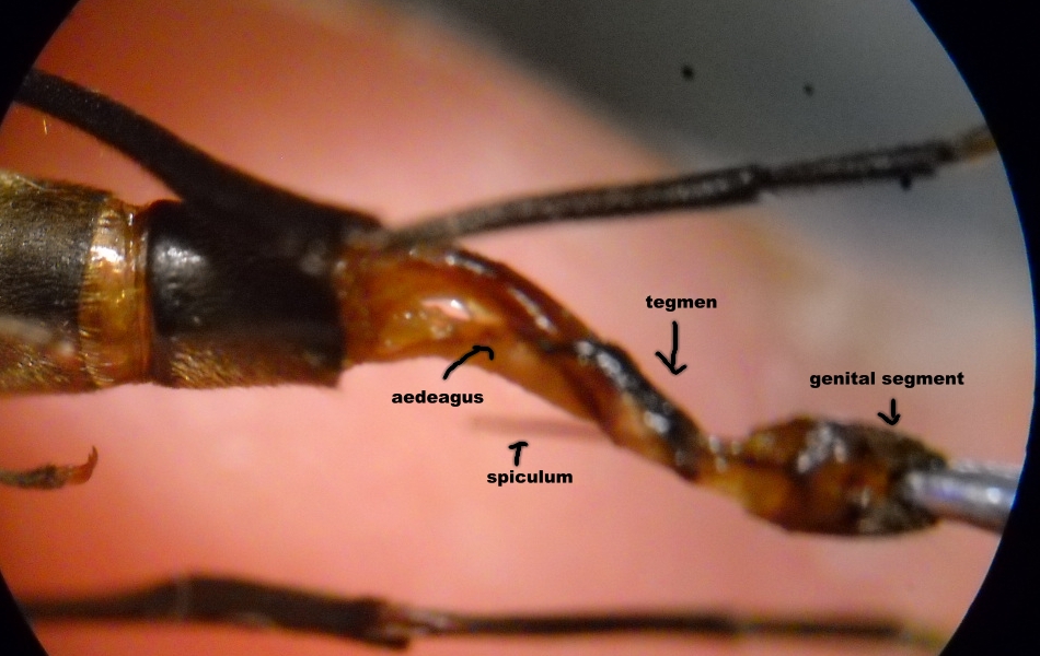
Posted - 16/03/2014 : 12:43:49
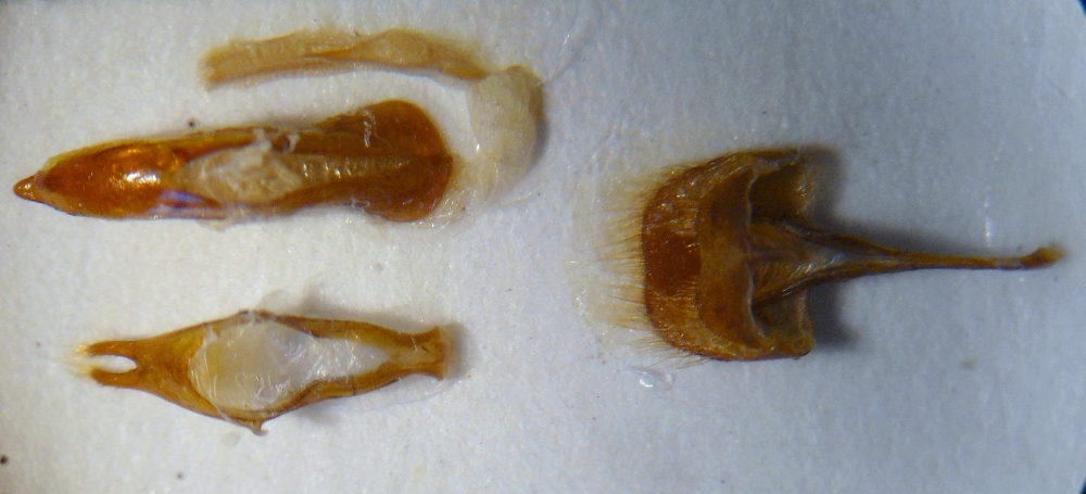
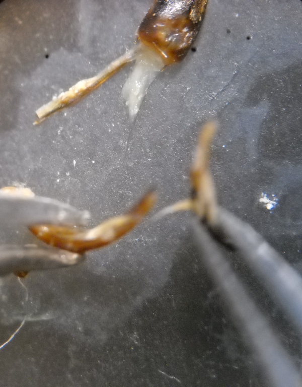
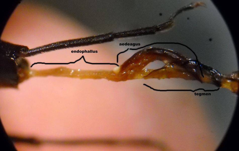
Now the three pieces are separated.
The tegmen is still covered with the rests of the membranes, which should be eliminated.
For a simple preparation, the pieces can be glued on a label, which will be pinned in the same pin of the (mutilated ) insect.
Here it is a simple example (the cleaning of the aedeagus was not perfect... ): ):
 |
| Francesco |
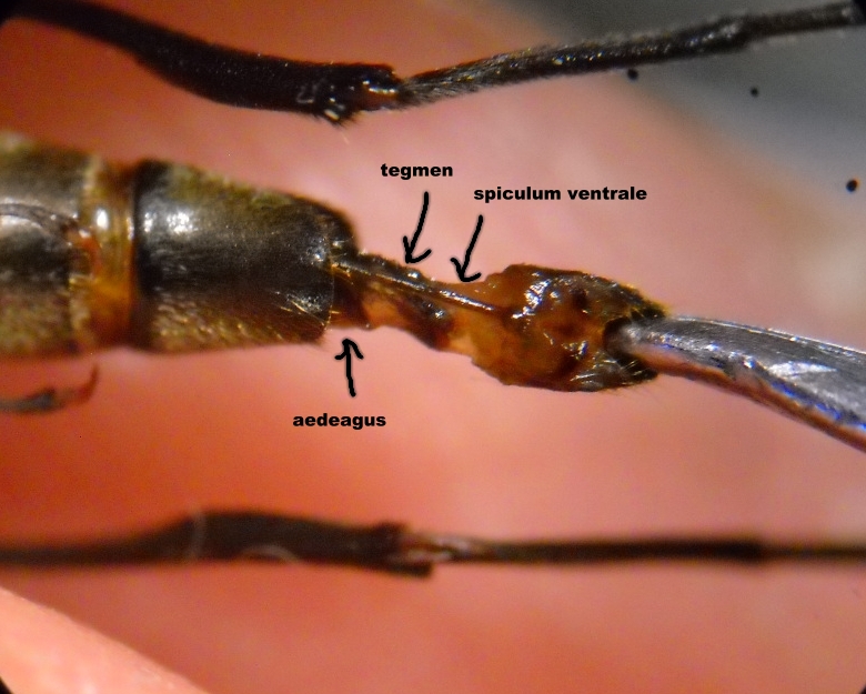
Posted - 16/03/2014 : 12:33:37
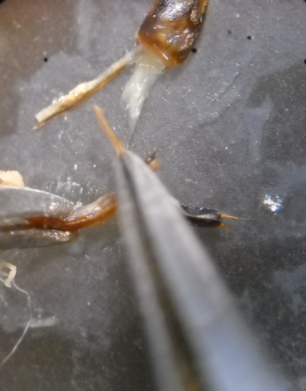
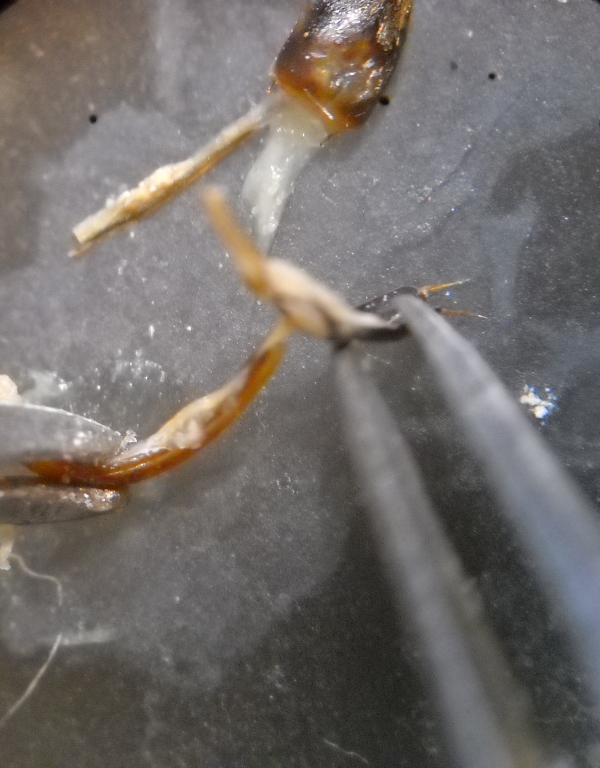
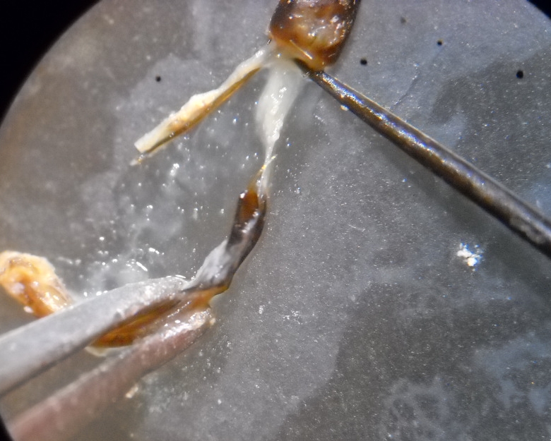
Separation of the tegmen.
The tegmen is linked laterally to the aedeagus so that the pieces act as a pair of scissors.
The superior part of the adeagus is linked to the tegmen with another membrane, which can be very resistant in large genitalia.
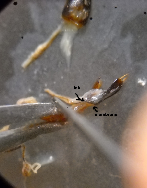
Now, holding the aedeagus with a forceps, take the basal part of the tegmen and (gently !) pull the tegmen outside.
If it makes resistance (this means that the membrane is too strong, dispose a second forceps like a bridge so that to pull contemporaneously both anterior and posterior part of the tegmen.
  |
| Francesco |
Posted - 15/03/2014 : 12:15:01
Step. 5: Cleaning
Before to begin this part, I suggest to plug the pieces in a vial with water and to hydrate them for some hours.
The membranes will become more easy to be separated.
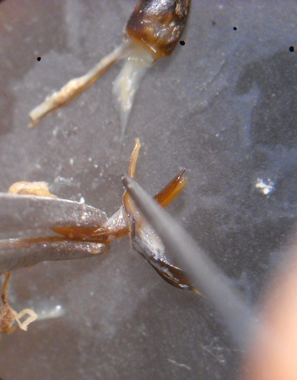
Separation of the genital segment.
Holding (gently!) the aedeagus with the forceps, put a pin transversely and pull the segment outside.
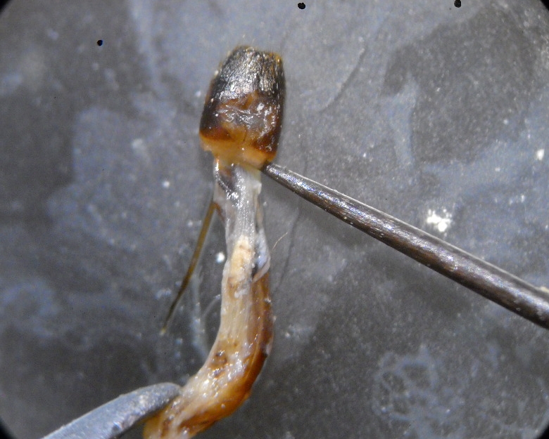
Now, the segment can be glued on a label. |
| Francesco |
Posted - 15/03/2014 : 12:14:15
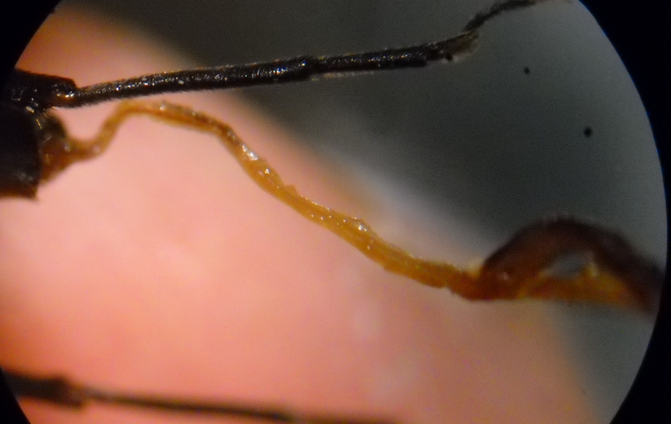
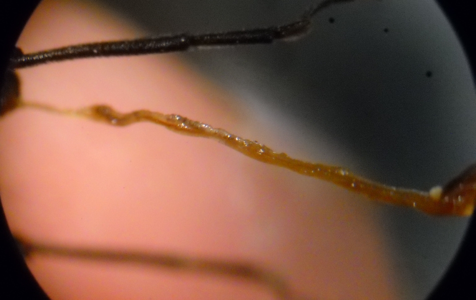
Here the pieces on the microscope tablet:
 |
| Francesco |
Posted - 15/03/2014 : 12:02:00
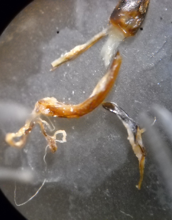
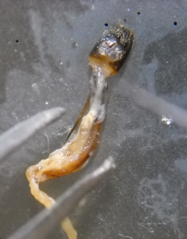
4. Step.
With the forceps pull gently the segment outside.
Normally, the membranes linking this segment with the previous ones should elongate.
If it makes too much resistance, then the insect is not sufficiently hydrated: hydrate it more.
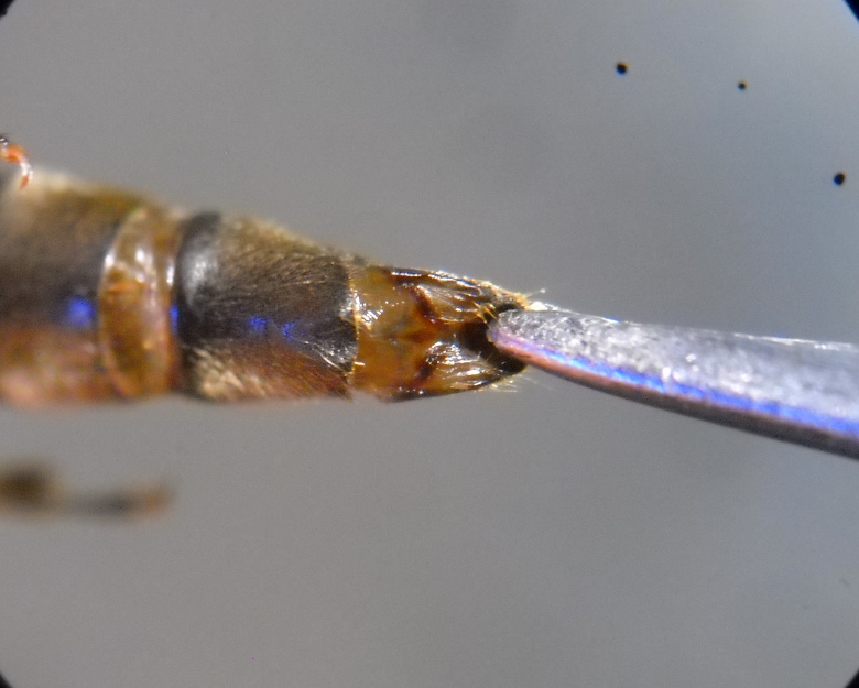
You should start seeing the aedeagus...
Pull gently!
You do not have to forget the endophallus!
to the end!
 |
| Francesco |
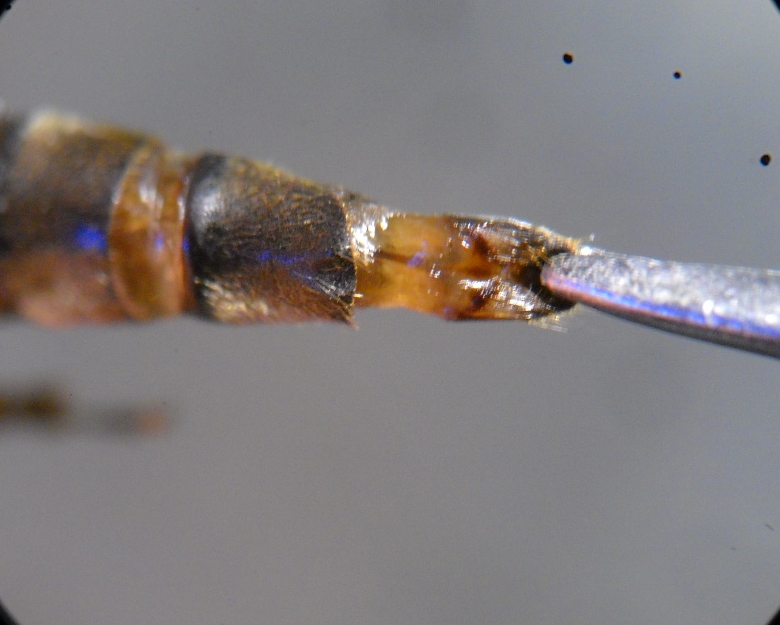
Posted - 15/03/2014 : 11:55:50
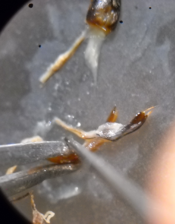
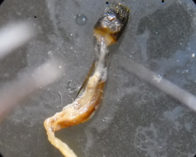
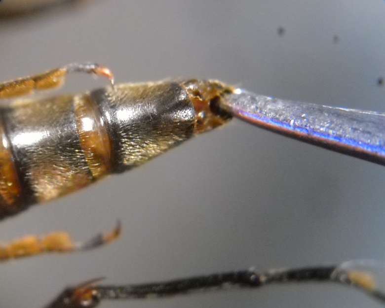
3. Step.
Holding the beetle with the left hand, take the genital segment with a pointed forceps.
Pay attention that the aedeagus might be located close to this segment. In this case, it looks like a yellow pointed lobe.
You must take only the genital segment!
 |
| Francesco |
Posted - 15/03/2014 : 11:43:03
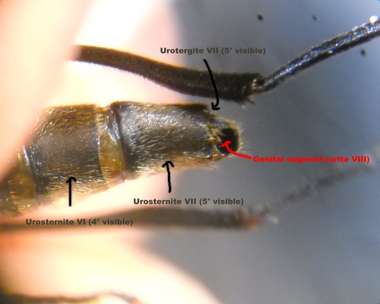
2. Step Individuation of the genital segment (VIII)
Each abdomen is constituted by dorsal and ventral segments (urites or ventrites).
The dorsal urites are called urotergites, the ventral ones urosternites.
Cerambycids normally show 5 visible urosternites (actually the urosternites III-VII).
The urite VIII is modified, apparently resulting as a single tergite.
Well, this step consists in the opening of the anal aperture and individuate this segment located close to the dorsal part of the abdomen.
 |
| Francesco |
Posted - 15/03/2014 : 11:24:16
1 Step. - IMPORTANT!
I suggest to start with banal, freshly killed, species.
Dried specimens must be hydrated as much as possible in order to dispose of flexible membranes.
But, specimens killed in alcohol (or formalin) and then dried must be avoided, since muscles and membranes are definitely fixed (baked!) and they cannot be sufficiently hydrated again.
In this case, the preparation will always be very difficult or nearly impossible.
|